HOCKS WITH HATS

The hock is a complex structure with many bones, tendons, ligaments and bursae. Today we are talking about the type of swelling that is commonly known as ‘capped hock’ and forms a round swelling, like a baseball cap, on top of the bone termed the calcaneus, the point of the hock, which is equivalent to the heel of our foot.


Tendons, equivalent to the Archilles tendon, attach at this point, while another (the superficial digital flexor tendon), travels over the point of the hock and continues down the limb.

To provide cushioning to these tendons there are fluid filled bursae between these tendons and the skin. 

A capped hock occurs when there is swelling of the subcutaneous calcaneal bursa and is typically the result of trauma such as kicking a fixed object. However, wounds in this region can also provide a point of entry for bacteria and in 40% of horses the superficial subcutaneous calcaneal bursa joins up with the deeper intertendinous calcaneal bursa allowing infection to spread to deeper tissues. 

Therefore, wounds in this region are an emergency. Injury to the tendons or fracture of the calcaneus can also cause swelling in this region so radiographs and ultrasound are sometimes necessary to know the best course of treatment.
Kissing Spines

This disease is known as Dorsal Spinous Process Impingement.


It causes back pain in horses which presents as reluctance to work and go forwards, bucking under saddle, muscular pain, or grumpy behaviour.


The cause is due to the spinous processes in the back, most commonly of the thoracic vertebrae 14-16, causing pain and irritation by being too close together.


The cause of the spinal process proximity can be due to spinal conformation, poor muscular development, lameness or back pain for another reason.

Poor core development also plays a large role in the onset of this disease.


How do we diagnose this problem?

It is a very complicated disease process, so come and see us out our Winter Woollies Seminars this year and I will be spending 15 minutes covering this very important topic.


Tuesday 11th July: Alexandra

Wednesday 12th July: Arrowtown

Thursday 13th July: Wanaka


Booking information will be emailed out soon.

June Dental Facts

We couldn’t start dental month and not put some fun Friday facts up about teeth!


How many teeth do horses have?

Up to 44 teeth. But most horses only have 33.


How are horse molars different to human molars?

Horses are born with very long teeth that hide in the skull. As they age, they continually erupt, and therefore their total length gets shorter and shorter, kind of like a pencil as its sharpened and used. Older horses’ teeth are therefore only anchored into the jaw by a small amount. They are very at risk of becoming loose and therefore painful.


Why do we rasp points from teeth and not just cut them off?

Horses’ teeth have multiple shallow pulp cavities containing nerves. Horses’ teeth are also strong to some forces, but brittle to others. By rasping, especially the use of power tools, we minimise the physical trauma to the tooth and the risk of fracture. Cutting teeth is VERY DANGEROUS. There is no way of knowing how close the pulp cavity is. Exposure of the pulp would be extremely painful for a horse.


Should tartar be removed from canine teeth?

YES… commonly fractures hide underneath the tartar which are very painful. If you can safely flick this tartar off with your nail, then you should.


SMILE… have a great day!

Winter Weight Loss

Some of our clients are finding that their horses have been losing weight over the last 4-6 weeks.


Some tips to ensure your horses stay healthy over the Winter months:

Oral health check prior to winter; ensures they are getting the most energy from their feed.


Shelter: this can be in the form of a physical shelter or a rug


Hay: Ad lib hay is ideal for any equine patient. Ensure each horse has 1.5% of their body weight in quality fiber (10cm long) per day. Please weigh your slabs of hay but often 1 slab of small bale hay is approx. 2KG. CAUTION, some horses will eat more than they need to stay at a safe weight.


Bullying: when feeding more than one horse, please be mindful of how you present them their hay. Some horses can be chased off their share of the hay if their paddock mate is more dominant.


Hard feed: consider using hard feed this winter to increase calories if your horse is losing weight, but do note, winter is the hardest time of year to put weight on horses as the additional calories cannot often be gained from grass. The aim would be to prevent unwanted weight loss.


Gastric Ulceration: this is always something to consider in any horse that is not gaining weight or is a picky eater. Do remember, it is ideal that horses can graze 24/7. BUT CAUTION, we do not want to have to diet overweight horses in the spring. So please monitor for weight gain.

Wormy Worries

For all of my clients that know me, I do love the topic of Equine Parasitology aka worms in horses.


I AM VERY WORRIED…. so, you should be too!


Last year at my seminars I discussed the concerns we have with our wormers and that they are no longer working the way they used. I advised all of my clients to be very careful when bringing new horses onto the property to ensure they do not bring a population of resistant worms.

Sadly, this season we have seen multiple horses with worms that are resistant to 2 different types of wormers, leaving us with minimal options going forwards.

A quick note: Resistance is NOT being seen in the way you would think. The wormers are reducing the egg output of the horse’s poo to zero. BUT not for long enough. The eggs are re-appearing much more quickly. This is an early warning sign.


What can you do?

PLEASE STOP WORMING unnecessarily.

A lot of out wormers are lasting 18-24 weeks before a positive egg count is seen. This means that horses may only need to be wormed twice a year, or even less if possible.

To all clients that worm at the change of the seasons or 3 times a year… PLEASE STOP. This could be very dangerous.


What do you need to do?

Get advice from myself or an Equine Vet that has an interest in horse worms.

The aim is to keep your paddocks clean by reducing egg output in the poo, and worm only when the egg count indicates it.


PLEASE make me happy and stop over worming.


Another DANGER:

Stop using any wormer that contains Ivermectin. If you want more information or to know why, get in touch.

READ the ingredients of your wormer: Cross out the word ‘Praziquantel’. And if there is only 1 more ingredient…. Do not buy it!!! Or throw it in the BIN!!


PLEASE let’s worm responsibly. We cannot turn back the clock.

I cannot name the wormers not to use, but if you go by my above advice, it will be easy to work this out yourself.


Thank you for helping save horses lives across NZ.

Temporary Fencing Risks

The injuries that commonly occur with electric horse tape are deep lacerations, and thermal injuries. Initially the injury may only look small, however the friction trauma that occurs when the tape is pulled across the leg, heats up the underlying tissues, and as time passes, the heated tissue begins to die off. As the tissues dies off there is risk of infection.

Some injuries can affect the deeper tissues which includes the underlying tendons or joints.


If you horse has a fencing injury, firstly, always get some advice. Send us a photo and we can help advise on the best next step.

Next, ensure the injured site is monitored carefully.

Apply appropriate topical treatments; this could include a spray, cream or a bandage.


What to look out for:

Increased swelling

Lameness

Pain on palpation of the area


What we can do:

Manage infection to the tissues

Check for damage to the deeper tissues

Pick the best treatment plan to get your horse recovered as fast as possible.


Our patient showed no pain or lameness, and minimal swelling for the first 3 weeks. Then he became acutely lame and swollen associated with infection. We treated him at our facility, and an ultrasound confirmed no underlying damage to the tendons. Treatments with sodium iodide have helped to reduce the scar tissue which is inevitable with a circumferential wound like this one.

Colic

 My phone ringing has increased, and my sleep has been decreased. Why?


There have been quite a few cases of colic recently!


This is most commonly due to dietary changes at this time of year.  (This time of year has also increased the number of rodents trying to move in – see photo of Becci in our feed bins)!

Any time there is a change in diet, there is a big risk of colic. Colic is the expression of abdominal pain; it is not a diagnosis of the cause.


What has changed in your horse’s diet to cause colic?

Increased grass growth causing changes in hind gut microflora resulting in hind gut inflammation.

Secondarily, we have seen gas build up in the bowel, and on occasion, movement of the bowel to an abnormal position.  If the abnormally positioned bowel doesn’t right itself, then surgical correction is required.


Best treatment: PREVENTION.

Try to limit your horse’s intake of fast-growing grass.

You can bring them into a smaller area and feed hay whilst your autumn grass grows to >10cm in height. Then it can be fed out as winter fibre and hay replacement.


Next, be proactive and consider using Equishure.

Equishure is like cooking bicarbonate soda. However, it is specially formulated so that it is not absorbed throughout the gastrointestinal tract until it gets to the hind gut. This stops the acid produced by the changed microflora and helps prevent pain, discomfort and irritability.

These can cause signs of:

Colic

Irritable behaviour

Problems under saddle


Please always treat colic as an emergency and get in touch as soon as you notice symptoms such as:

Reduced appetite

Pawing at the ground/stomach

Excessive rolling and/or sweating

Restlessness

Flat Back Feet

This week we discovered multiple horses with flat angles to their pedal bone in the hind feet. Most horses would measure an angle of 3-5 degrees between the ground/shoe surface and the solar surface of the pedal bone. The difference depends on breed and conformation. The importance is related to good biomechanics.


Solar angles of negative or up to 2 positive degrees in the hind feet can cause pain via the following pathways:

Inflammation and damage to the soft tissue structures of the back of the foot.

Inflammation of the hock, especially those with pre-existing arthritis.

Pain over the back, gluteal muscles and hamstrings.


Horses can present with the following clinical signs:

Poor performance/unwilling to go forward

Bucking/behavioural changes

Bilateral hindlimb lameness

Hock soreness

‘Bull nose’ appearance to the toes of the back feet


Treatment:

This depends on the time of year it is diagnosed but could include:

Aggressive trimming of the sole at the toe

A heart bar shoe +/- wedge

A wedge pad with packing material or equithane


Always consider this when you are concerned regarding poor performance.

Fun Facts

Our facts today related back to the times when horses were introduced to NZ. At this time, they were key workers for farming, development, transport, and for some people, pleasure riding:


Introduction of Horses to NZ:

The first horses in NZ were a stallion and two mares brought from Australia.

They arrived in the Bay of Islands on 22nd December 1814.

By 1900 there were > 260,000 horses in NZ.

In 1911, the horse population reached its peak at 404,284 ( 1 horse per 3 people)

In 2004, horse numbers reduced to 76,918.


Horse type:

Draught horses for working were the main horse type for hauling and ploughing until 1950’s.

When mechanical engines were introduced, their power was measured in terms of horsepower: The power to life 15 tonnes, one foot high, for 1 minute.

The Scottish Clydesdale was the main draught breed in NZ.

The first Coach service to travel across the country started in the 1860’s: Reportedly it was bumpy!!

The Hackneys were imported in 1880’s for carriage work.

Next came the import of Arabians, Thoroughbreds and Standardbreds.

In the recent years, American breeds have also become popular.


Feeding:

Horses at as much as eight men or four sheep: large amounts of valuable farmland were taken up in growing oats for them.


Wow, how much times have changed 😉

Bandaging

Just a couple of photos this week to assist in bandaging technique for lower limb wounds.

We have seen a few fence injuries recently that require bandaging. Bandaging is a great way to improve wound healing by promoting granulation tissue which prevents infection. In addition, it then promotes new skin growth which inhibits proud flesh formation.

NOTE:

A layered bandage is required to avoid painful bandage sores. See photo diary.

This technique does not apply to the Carpus (knee) or Hock.

Bandages over mobile joints need a different technique.

1: These are examples of a primary layer to a bandage. These dressings are non-stick and should be held on by a conforming padding such as softban seen in the picture.

 2: This picture shows the softban holding the non-stick dressing in place.

3: Next is the secondary layer to the bandage which consists of padding. Padding helps to absorb wound discharge but also reduces the risk of bandage sores. This padding can be of different thicknesses depending on requirement.

 4: The last layer is the tertiary bandage layer and consists of vetwrap. This compresses the padding and can be used under different pressures for desired result.


5: The Elastoplast is used to assist in compression, but also to prevent foreign material from entering the top and bottom of the bandage. It also acts as a shower resistant layer.

Eye See You!

Over the last few weeks, we have seen many emergencies due to traumatic injuries of the eye and more specifically, the cornea.

The cornea is the clear/see through structure at the front of the eye. It contains lots of nerve endings and hence any trauma is extremely painful.

In addition, it has its own normal microflora, but when there is damage to the cornea, opportunistic bacteria can take hold and cause an infection.

When bad bacteria multiply in a horse’s eye, it causes a dramatic response from the horse. An imbalance can happen in the tear film between enzymes that promote degradation of proteins and enzymes that inhibit this breakdown. As a result, the cornea can start to be dissolved. We say it is ‘melting’. The scientific word for this is ‘Keratomalacia’.


Take home message:

Treat every painful, swollen or tearing eye as an emergency.

Antibiotics are almost always used topically when waiting for a corneal trauma to heal.

Do not wait to call for advice.

Once bacteria trigger an increase of these destructive enzymes, there is limited time to react before the cornea can perforate (burst) and the horse requires eye removal.

Canine Teeth/Extraction

Dental examinations are extremely important to pick up on any underlying painful conditions in your horse’s mouth. Do not rely on behavioural changes as an indicator for oral discomfort as often no change is reported.


This post reminds us that any time after a dental procedure, a tooth can be broken. This canine tooth shown in the photos, fractured likely due to him catching his tooth under something. This happened only a few months after his annual dental check.

If our client had not observed his demeanor change, then he could have had this painful condition for a further 8 months.


The canine tooth is actually a very large tooth, however the clinical crown (the bit we can see), is only a small portion of the size of the canine tooth under the gum line. They are also ‘Cashew Nut’ shaped, which makes extraction more difficult. The bone on the outside surface of the tooth needs to be removed to enable the tooth to be removed without leaving the root inside.

Many clients incorrectly believe that mares do not get canine. They do; however, they are more common in male horses.


See you all in June for our annual Dental Special month where we put a big focus on oral health.

Right Dorsal Colitis

We use Bute (phenylbutazone) a lot in horses, and it is a great drug for pain relief – but it does have some side effects that we need to be aware of. One of these is right dorsal colitis (RDC).

The right dorsal colon is the last segment of the large intestine, and it is only this segment of the large colon that is affected by Bute.

Why this region of the large intestine? That is a question that has baffled vets for a long time, but recent research is pulling together some pieces of the puzzle and are finding that this segment of the large intestine uses unique secretions to protect the lining of the intestine from the volatile fatty acids that are produced in the large intestine.

Bute can interfere with this protective secretion, and this can result in severe ulceration and subsequent protein loss and diarrhea which can be fatal.


So, Bute does have some downsides and should only be used under veterinary guidance. When your horse is on Bute pay attention to your horse’s manure and if they produce less than 10 piles a day or if it’s starting to get like a cow-pat consistency, give us a call.

Sheath Cleaning

The sheath is the blind ending sac that contains the penis.

There is no requirement to clean this unless it becomes dirty.

How do I know if the sheath needs cleaning?

To check if the sheath is dirty, you just place a gloved hand up into the sheath on the belly side of the penis and look for a moist paste on your hand when you bring your glove out.

If this is present, then the sheath needs a clean.

Be careful, some horses do not like this being performed.


Cleaning the sheath:

Use purpose made sheath cleaners only, and some warm water with cotton wool to remove the excess material.

Don’t forget the penis:

What’s most important is that a penis health check is performed at the same time: This would include checking for a bean, examining the entire penis skin to ensure there is no early signs of cancer. A pink penis would be particularly at risk.

Often to do this entire genitalia area health check, your horse would need sedating to relax the cremaster muscle that is responsible for keeping the penis up into the sheath.

We recommend during your yearly dental check that we perform this procedure for you; this is a free of charge service if performed during your annual dental.

The picture shows a wart like growth on the inside of the sheath. This should be monitored carefully as this could potentially be a cancerous change.

Weird and Wacky Walking

There are many different reasons why our horses might walk a bit odd – usually it is because they are lame but occasionally it is due to nerve dysfunction. Shivers and stringhalt are two conditions that are easily confused as they both cause the hindlimb to be lifted high up to the belly (hyperflexion) as the horse walks.


With Stringhalt, the abnormality is most severe at a walk and the hindlimb snaps sharply up to the belly then forcefully stomps on the ground.  Stringhalt is caused by a toxin in the plant catsear (also known as false dandelion or flatweed). We do have catsear in Central Otago and like its name suggests it looks very similar to dandelion. The main difference is that catsear has hairy leaves and a stem that branches to produce multiple flowers while dandelion produces a single unbranched stem. What’s the best way to get rid of catsear? Sheep and cattle can eat it without any issues, but if that isn’t an option, herbicides can be used.


Shivers also causes hindlimb hyperflexion but, unlike Stringhalt, is most severe when the horse is being backed up. Also, in addition to being held upz, the limb is often held out from the body and trembles. Shivers is caused by damage to special nerve cells in the brain and is a slowly progressive disease. Draft horses are most commonly affected but other breeds can get this disease too.

Sleeping in a Sling

Sleeping in a sling:

What is the Stay Apparatus?

This describes a group of ligaments, tendons and muscles strategically placed within the skeleton of the horse. There is a complex network of ligaments which act like tension bands and enhance stability. Please see the images attached.


The function of the Stay Apparatus:

Its main function is to ‘fix’ the horses’ legs in a straight position allowing minimal muscular effort when standing.


What is the job of the Stay Apparatus?

It minimises muscular fatigue and also allows horses to enter lighter stages of sleep without falling over.

Please note, horses still need to lie down daily for a deep sleep.

When is the Stay Apparatus activated?

Relaxation of muscles causes the engagement of the stay apparatus.


Some more details:

The stay apparatus can be separated into the lower limb and upper limb.

Lower limb: This is the suspensory apparatus and is identical in the fore and hind limb.

Upper Limb:

The forelimb and hindlimb are different. Please see the attached diagram.

Additional and unique features within the hind limb:

It has a locking stifle system and also a Reciprocal Apparatus.

1: The stifle of the horse locks when the patellar is hooked over the bony ridge on the inside of the femur. Active muscular contraction is required to lift the patellar up to release the lock.

2: The Reciprocal Apparatus is a modification of largely tendinous muscles, that act oppositely on either side of the tibia.

Common things that can go wrong with the special features of the hind limb?

1: Unwanted patellar locking. This can be due to many reasons but includes poor muscle tone in the quads, inflammation or pain within the stifle joint, or an underlying lameness.

2: Damage to the Reciprocal apparatus. This is commonly a traumatic injury.


A clinical case this week:

This week we saw a poor girl that had broken the tight band of the reciprocal apparatus. She ruptured her Peroneus Tertius.

This happens as a result of over-extension of the leg. She is able to flex the stifle when she moves but the hock remains in extension. We wish her all the best in her recovery which will include lots of rest and relaxation.


Get in touch if you have any questions. Let us know if you need help with yarding your horses during recovery from musculoskeletal skeletal disorders. We have specially designed small paddocks for this.

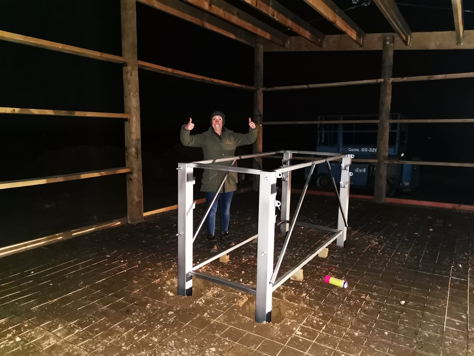
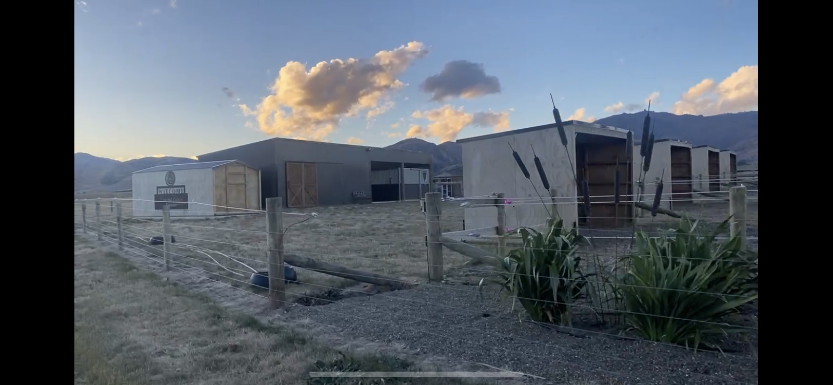
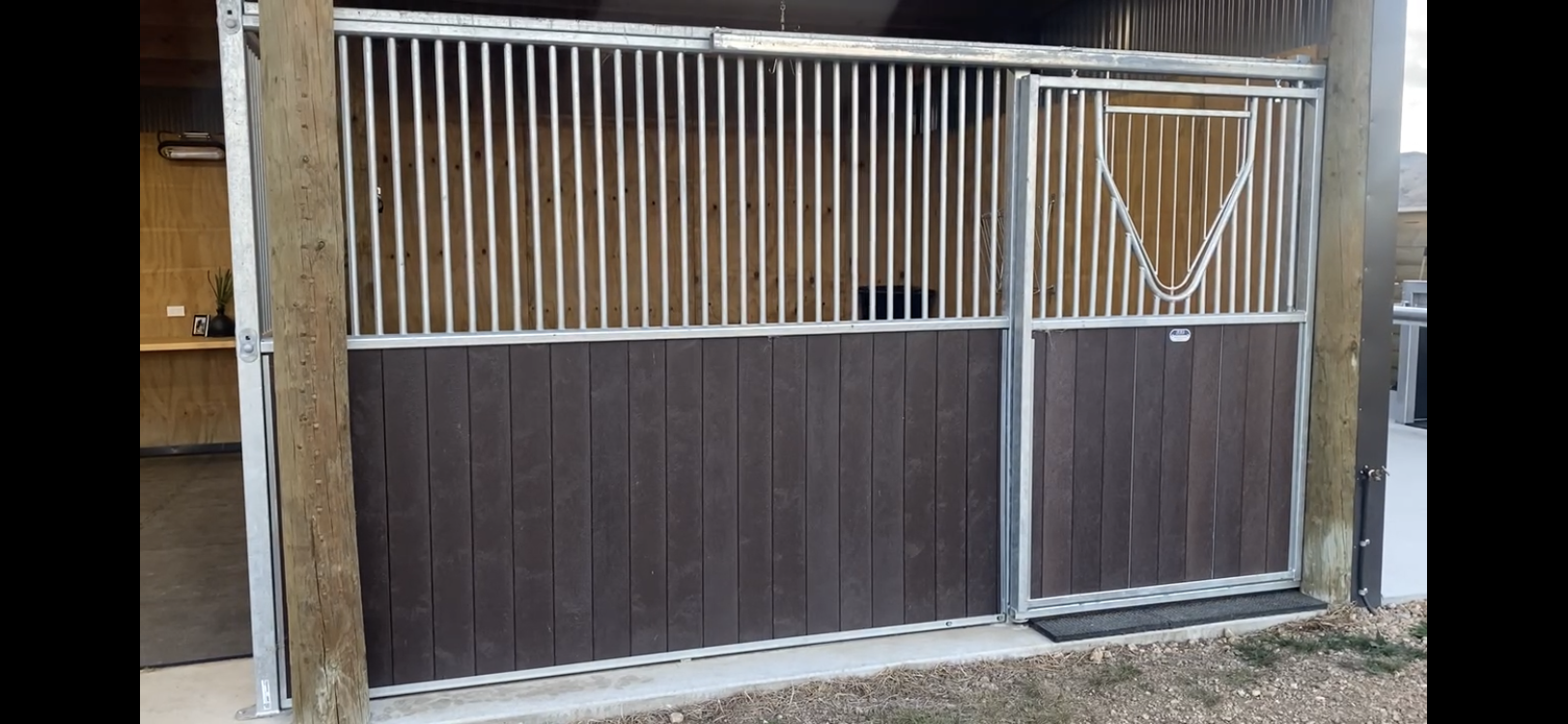
Building Blog

Fun Facts

Fun Facts:


  • What is different about blue-eyed horses?

Commonly they have Iris Hypoplasia. It can be seen as a bulge in the iris. Changes inside the eye should always be checked, but this change is often normal.


  • Do mares have canine teeth?

Yes, they can do, and it is very normal if they do.


  • How many nostrils do horses have?

Technically they have 4. Each main nostril has an additional blind ending sac on the inside which we call a false nostril.


  • Why are foot abscesses so painful?

The swelling that occurs due to the infection has nowhere to go as it is confined by the hoof capsule. The resulting pressure is very painful. Often even anti-inflammatories do not help. The pressure must be released. Get on the phone to your farrier first 😉 especially if it’s a Friday night hehe!


Pedal Bone Fractures

Pedal bone fractures are relatively common. The cause of them is variable, but we most commonly see them due to trauma (such as kicking a wall), or in horses with flat feet and thin soles which are galloped on hard ground.

Clinical signs of a pedal bone fracture:

‘Foot Soreness’ or ‘Acute Lameness’

Hoof tester response may be positive or negative.

Inflammation of the foot noted with increased digital pulses

Diagnosis:

Radiographic examination: The observation of a fracture line confirms the diagnosis.

Playdoh is used to help to remove superimposition of normal lines on the foot.

Sometimes if the two fracture pieces have not separated, then a repeat radiographic examination is needed 2 weeks later to be able to see the fracture line. The fracture is more evident as this stage due to remodelling of the fracture ends during the healing process.

Treatment:

If there is no involvement of the joint surface, a bar shoe with or without a foot cast is used.

Surgery is not needed.

Generally pedal bone fractures heal very well with a remedial shoe.

Fun fact: Pedal Bone fractures can heal but leave a permanent radiographically evident line for generations

Why?

They can heal by a fibrous union!

Our patients are very lucky that we are able to take radiographs in the field to give us a ‘horse side’ diagnosis to provide the best possible care for our patients.

Chemotherapy

With all the wind and rain recently, we are very glad to be able to perform chemotherapy inside the hospital.


There are many different options for treating skin tumours depending on the type of tumour that is present, and the location of the tumour.


This patient has an advanced ocular sarcoid of a mixed type. The treatment being used is an emulsion of slow-release chemotherapy drug injected throughout the skin tumour.


I will explain in more detail, the use of spinal needles and local anaesthetic for this procedure:


Spinal needles-

Spinal needles are normally used for injecting fluid into the central nervous system. Here they are being used to prevent tumour cells from getting stuck inside the lumen of the needle when they introduced into the dense highly cellular tumour . The spinal needles have a central piece of metal that prevents the needle from becoming clogged up. When the central piece is removed, a tunnel is left for the chemotherapy treatment to be left in.


Local anaesthetic-

Local anaesthetic is used to ensure our patient cannot feel the surgical procedure. There are different types of anaesthetic, but they all work by preventing nerves that monitor pain from firing and sending messages to the brain that it is painful.